Optical Projection Tomography Scanner
Optical Projection Tomography Scanner
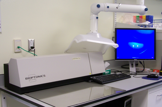
The OPT is a device that collects a series of 2D projection images by rotating a specimen 360 degrees around its central axis. These images are reconstructed into a 3D volume that can be re-sliced in any direction. The resolution of the scanner is best suited for macroscopic analysis such as wholemount gene expression or whole antibody staining of embryos. The maximum specimen size is approximately 10mm wide x 10mm high x 15mm long. The OPT employs two types of illumination: bright field and fluorescence. Flourescence illumination consists of three available channels: texas red, FITC green, and a wider spectrum green channel. It is essential to achieve optical clarity in the specimens, and therefore careful preparation of samples is required prior to scanning. Sample preparation can be performed outside of the Centre for High-Throughput Phenogenomics in the user’s home lab, allowing for ease of monitoring. Sample preparation guidelines can be provided during training on the OPT. A major advantage to the OPT is the rapid acquisition time, which is approximately 3-5 minutes per channel depending on the resolution. Furthermore, reconstruction can be performed on one sample while another scan is running, or if preferred, reconstruction of samples in batches can be performed at a time separate of acquisition. Following reconstruction, volumetric measurements of segmented areas are possible using software provided on the computer terminal. Additionally, reconstructed images can be saved in many file formats, allowing the user to retrieve their data and perform the analysis in their home lab if preferred.


MORE INFORMATION
For more information about the capabilities and use of this machine, please send an e-mail.
BOOKING CALENDAR
Click here to view the booking calendar for the Optical Projection Tomography Scanner…